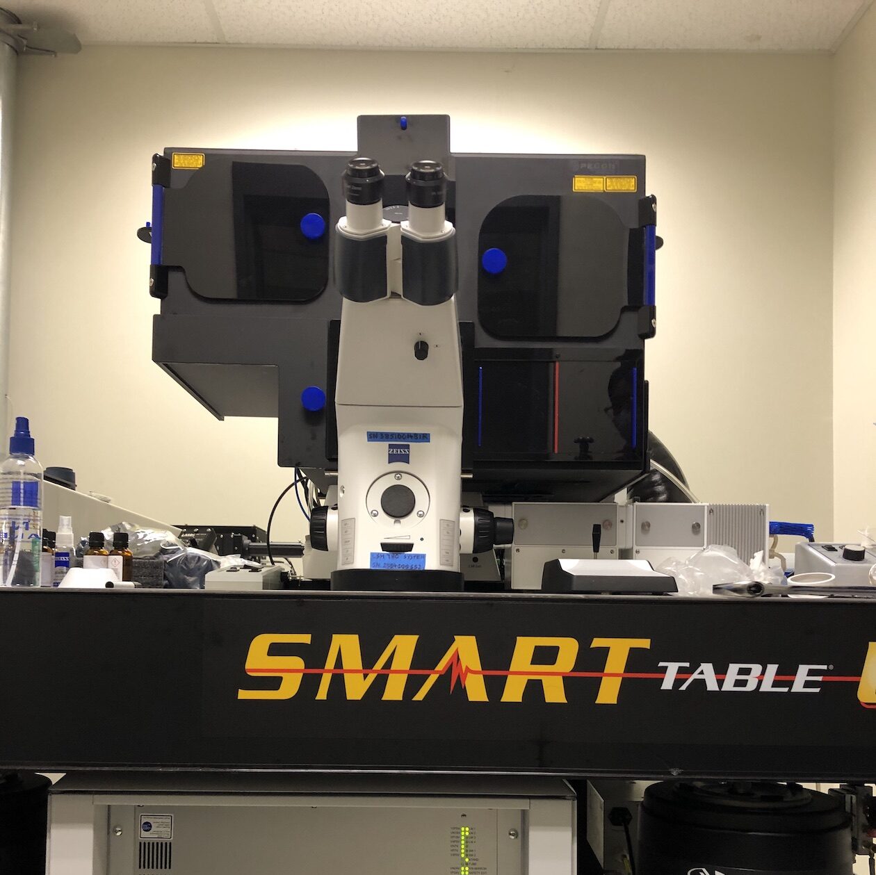
Zeiss LSM 780 NLO

Overview
NeuraCell is proud to offer the advanced imaging capabilities of a new Zeiss LSM 780 NLO spectral confocal & multiphoton microscope. Confocal microscopy employs optical sectioning to exclude out-of-focus background noise and to generate 3-D image stacks, and is ideally suited to imaging thick, fluorescently labeled tissue specimens ideal for tissue specimens 50+ microns thick. Even with cell culture monolayers, confocal microscopy provides detailed 3-D subcellular localization. Multiphoton microscopy facilitates imaging even deeper into thick specimens, and does so with reduced photobleaching and phototoxicity, making it ideally suited to longer-term, live imaging studies.
The NeuraCell Zeiss LSM 780 NLO boasts an environmental enclosure for long-term live imaging studies and extremely sensitive state-of-the-art GaAsP detectors for standard confocal, spectral confocal and multiphoton imaging. The GaAsP detectors may be operated in photon counting mode, enabling sensitive fluorescence correlation and cross-correlation spectroscopy (FCS & FCCS) experiments. Spectral confocal permits separation of overlapping fluorescence emission spectra especially useful for specimens tagged with multiple (5+) markers. Non-descanned detectors (NDDs) for optimal multiphoton sensitivity include both conventional and GaAsP detectors, and can be configured in both reflected and transmitted modes for applications like second harmonic generation (SHG) imaging of e.g. unstained collagen fibers.
Optical Features
Multiphoton, nonlinear (NLO) excitation between 690-1050nm is provided by a Spectra Physics MaiTai DeepSee laser. Continuous wave (CW) lasers include:
- 405nm diode
- 458, 488, 514nm argon
- 543nm HeNe
- 633nm HeNe
Objective lenses suited to a wide range of applications are available:
- 10x/0.3 for navigation.
- 25x/0.8 multi-immersion for larger fields of view.
- LD 40x/0.6 long-distance, dry objective for plastic dishes & multiwell trays.
- 40x/1.3 water immersion ideal for FCS.
- 63x/1.4 oil immersion for high resolution subcellular detail.
Product or Service Inquiry
Have a question or interested in purchasing? We are happy to help!
Image Analysis
An extensive software suite covers everything from routine imaging to more demanding applications:
- 3-D image acquisition including timelapse, tiled montages, and/or multiple positions.
- 3-D image visualization, analysis and display including movie rendering, etc.
- Physiology module for analyzing cell & tissue dynamics.
- Fluorescence Recovery After Photobleaching (FRAP) experiments.
- Förster Resonance Energy Transfer (FRET) imaging and analysis.
- ROI-HDR module ideal for imaging dim features adjacent to strong signal (e.g. neurite processes projecting from neurospheres).
- Fluorescence Correlation & Cross-Correlation Spectroscopy (FCS & FCCS) for detecting single molecule dynamics.
- Macro recording and MultiTime macro for fully flexible experimental design.
All image analysis software modules are also made available on an offline computer station, free for use, to permit uninterrupted image collection.
